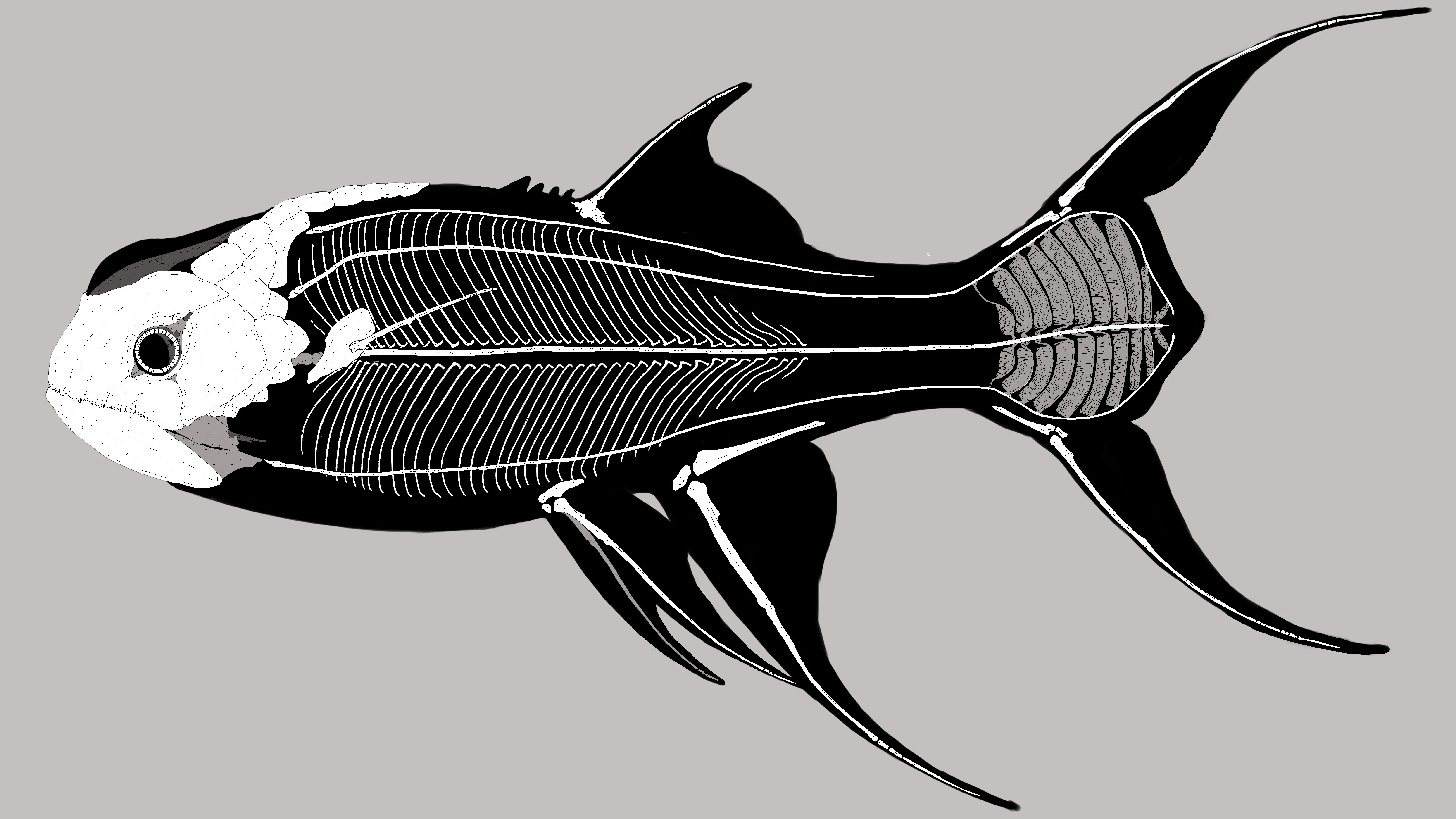
General skeletal anatomy
The general skeletal anatomy based on an adult specimen of Joannia equatorialis
Basic Information
Anatomy
Cranial Anatomy: The anatomy of a generic Eoichthyian, Pressionatatian or Eumuraenian amphibian is composed by the cranium and a jaw, both covered by armour-like bone structure. The cranium, underneath the armour, is made of light thin and porous bone with many fenestrae which function as anchoring points for the mandibular and neck musculature. The only cranial bones that are stronger and reinforced are the maxillary bones and the palatine. On the dorsal section of the cranium a bone stretches out, going way back near the base of the neck; this bone is the Sexual Hoplos and its main function is that of protecting the female's oviducts and egg sacs; this bone is present in both sexes, although only one of the two has the target organ the bone is meant to protect. On the underside of the cranium lies the Lanial Palatine, a modified bone structure with many, teeth-like jaggers extruding ventrally, towards the throat centre; this bone is one of the two used by the animal to cut up food and chew. The jaw, underneath the armour, is composed of a stronger bone, sturdier and heavier than most of the cranium; the bones themselves are very similar to Earth's reptile jawbones, with a Dentary and Angular and a Surangular bone but without any mandibular fenestrae. Coming out from the underside of the jaw there are three, thin, bones called the Lanial Ribs; the first two Lanial Ribs are arch-like bones anchoring a the Musculus adductor lanialis minor, the contraction and relaxation of said muscle move the Sphaera Lania (touched upon later) in a rotatory fashion, putting into motion the lanial apparatus and allowing the animal to chew food. The third, and largest, of the Lanial Ribs, anchors the Musculus adductor lanialis major and its contraction, upon the opening of the mouth, pushes the Sphaera Lania out of its socket and into the interior of the mouth, creating the crushing space necessary for the apparatus to work. The Sphaera Lania is a circular bone found inside the jaw of the animal; the surface of this bone is covered in jagged edges resembling teeth; these edges are extremely sharp and are constantly changed during an animals life, just like teeth in Earth's sharks; through the rotatory movement of the bone and the Lanial Palatine just above it, this one is able to crush and mince whatever the animal tries to ingest, be it flesh, plant matter or even bones. Armor: The armour covering the cranium and jaw is bony in nature, like the underlying bones it grows on; this armour, depending on the family or sometimes even genera can be Segmented, Fused or even Reabsorbed. Segmented Armor: There are various levels of armour segmentation visible mostly in Taratosomnian amphibians. The most segmented armors, like those of most advanced Víaiosmandibulidae (Draco basalis) are composed of three major sections; from snout to the post-temporal area in order are the Rostral section composed by the Frontal, Premaxillary and Lacrimal plate/s.
The Post-Orbital section composed of the Sclerotic plates (not always present especially in simpler segmentation), the Supraocular plate, the postorbital plates (up to five) and the Maxillary plate.
The last section is the Post-Temporal section which is composed By the post-Temporal plate, the auricle plates (up to three) and the Masseter plate. Fused Armor: The Fused armour is one of the most common and characteristic types of Cranial armour on the planet. A perfect example of Fused armor is the Eoichthyian amphibian Bigeye Spotted Limn; this armor type has most plates fused together, most commonly to the point where only two major plates remain on the whole cranial armor; these two plates are called the Fronto-Temporal plate and the Maxillo-Massenteric plate; In some genera the Lacrimal/s remain. Reabsorbed Armor: The Reabsorbed armour is in itself divided into two subcategories being the Pseudoabsorbed state and the Euabsorbed state. The Pseudoabsorbed armour is the most common of the two and can be exemplified in the cranial anatomy of amphibians such as the Blackfin Yung Zao; in this state, the cranial armour has fused into one single plate, simply called the Cranial plate; this structure is then absorbed by the underlying cranium into a thicker fused bone forming an akinetic skull; this absorption of the armour usually happens after the embryonic stage either in a prenatal condition or as an ontogenetical process during growth. The Euabsorbed state is best exemplified by the Esavelidae such as Common Flapper. In this more advanced state, the cranial armour has been adsorbed to such an extent where it is no longer distinguishable from the underlying bones, producing an akinetic cranium with normal density and porosity to the axial skeleton; in such craniums, it's still visible the distinction between the armour-born bone and the cranium bone at a phylogenetical level. Euabsorbed armours are absorbed during the embryonic stage of growth and as such are directly fused before birth to the skull. Axial Skeleton: Fronto-dorsal anatomy The frontal section of the dorsum is where most of the dorsal armour lies and the area in which the natal aculeus grows. The Natal Aculeus is a specialized plate forming from the base of the neck during the latest stages of growth before birth; this aculeus is essential to the animal as it is the only way it has to break the eggshell and free itself from it. Once the animal is born, the base of the aculeus gets slowly reabsorbed until the aculeus itself falls off. Some genera retain the aculeus to adulthood. The Dorsal armour, like its cranial counterpart, can be divided into Segmented, fused and reabsorbed types although this classification can quickly become confused as to the enormous variation it can have from genera to genera. The dorsal armour is overall rarer on the planet, with more amphibians having a reabsorbed one than ones with even a fused form. There are seven proposed methods to classify dorsal armours, none of which is universally accepted. The Body The main body structure of an Amphibian of Nijin-Konai is composed by six "Vertebral columns"; these columns are not composed of several vertebrae like Earth's vertebrates, though, they are closer to notochords, being composed of a single, long, flexible bone going down the length of the body. The Vertebral column I is central to the body and is in an analogous position to our fishes; this column is hollowed out and makes space for the vertebral cerebrum; it attaches to the cranium through the Atlantic suture, collocated posteriorly to the skull in what is the most caudal pointing bone of the cranium, the cervical cavity and magnum suture. In the cervical cavity, the cerebrum is located and it goes down the Column I for almost its entirety. The Vertebral column II and III are twins and are located dorsolaterally the I; these two Columns originate shortly after the caudal end of the cranium and go down to the Gill fan. These Columns act as an anchoring point for several muscles and as an anchorage point for the Upper Gill. The Vertebral Columns IV and V are located ventrolateral to the I and, much like the II and III, originate caudally to the cranium and go down to the Gill fan; these columns are also anchor points for muscles and the Lower Gill. The Vertebral Column VI is smaller than the others; it is located dorsally to the II and III and originated after the Sexual hoplos only to terminate middle way through the Gill tail; this column is the anchorage point of the dorsal musculature and also the anchoring point of the upper rib cage. Rib Cages There are two Rib Cages in an amphibian from the planet; these are classified as the Upper Ribcage and the Lower ribcage. The Upper ribcage contains the smaller part of the abdominal cavity and only protects the heart and upper intestines. The Lower ribcage offers protection to the bigger part of the abdominal cavity and protects the Stomach, the Filtration Sac and Lung and, in most cases, even the mouth of the intestine. Both these structures, through their paired morphology and curvature, create the perfect anchorage point for the Hypoaxial muscles of the animal. Gill Fan The Gill Fan is a specialized apparatus growing on the end of the tail of every amphibian of the planet; it is divided in Upper and Lower Fan, both having an Upper and Lower ridge, created by the musculature of the tail and the arch of the Vertebral columns II, III, IV and V. The bony structure of one Gill is composed by a pair of long and slender bones called Branchial Ribs, the Branchial ribs connect to the Vertebral column I by cartilage. Each of these ribs sustains a Branchial Osteomembraneum bone, a subrectangular to elliptical web of bony structures from which the respiratory canal attaches; the morphofunctional unit of these bones is the filtration coral, of which the entire Osteomembranaeum bone is made of. Through the constant movement of the Gill Tail, the Fan is able to take in water and process it for oxygen. Appendicular skeleton: In most of Nijin-Konai's amphibians, the limb bones tend to be fused together ina single rigid structure (outside of the Pes) so in this article about the general skeletal anatomy, I'll stick to describing this most common case. The scapula in the average amphibian is collocated laterally to the Column I and slightly caudal to the Atlantic suture; the scapula itself works very similarly to our vertebrate's with the only major difference being the pronounced scapular fossa in which the muscle governing the fin movement is located. The dorsal Hip bone opens up laterally to the body from the base of the dorsal fin; deep lateral processes help in the anchoring of the muscles and tendons used to move the dorsal fin. Genera that use the dorsal fin for active locomotion instead of steering have much bigger dorsal hip bones with deeper processes and fossa. The Anal Hip bone I is short, with a reduced pubis and ischium, while the Anal Hip bone II has a majorly enlarged Ischium for the attachment of the caudal muscles. If preset, the Caudal Hip bone I and II have elongated pubes pointing diagonally to the centre of the body; here the caudal muscles and tendons attach to give the animal the ability to open or close the caudal fins by actively pulling the hip bones towards the body centre. The limbs are composed of roughly the same bones as human legs; they have a Femur, Tibia and Fibula terminating in a Pes. Femur, Tibia and Fibula are fused together in a rigid bone, the membrum; the only bones that are still unfused are the Ankle, Heel and the single Metatarsal. Skeletal diagrams:
Remove these ads. Join the Worldbuilders Guild


Comments